


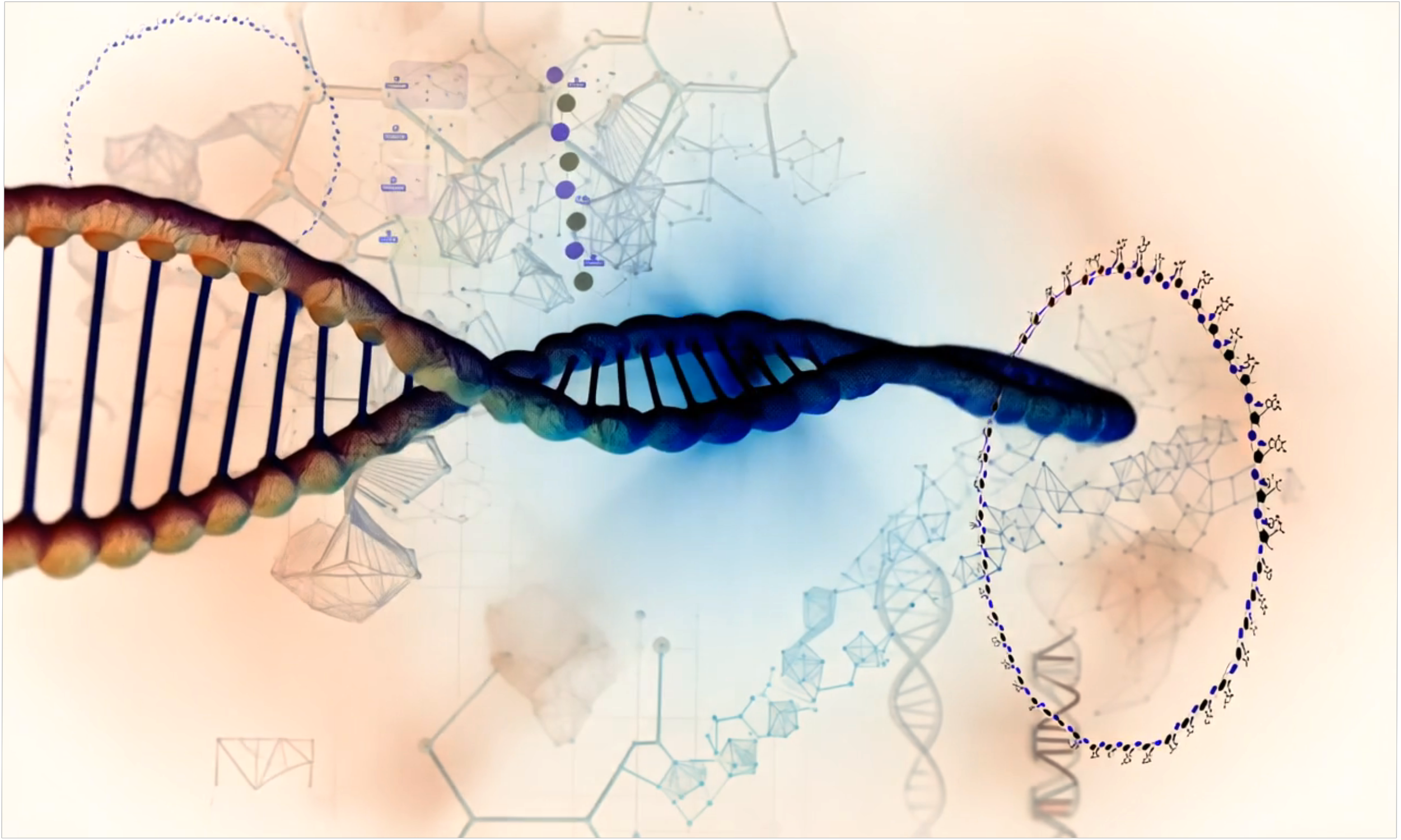
RNA Based Technologies
RNA technologies now span a broad and rapidly expanding landscape, encompassing therapeutic platforms, diagnostic platforms, gene editing and more.

In the past decade, RNA has transcended its traditional role as a passive intermediary between DNA and proteins to become a dynamic engineering substrate at the forefront of biotechnology, medicine, and synthetic biology. Unlike DNA, which encodes static genetic information, or proteins, which execute biochemical functions, RNA occupies a unique space: it can be rationally programmed to carry information, catalyze reactions, regulate cellular processes, sense environmental changes, and even assemble complex nanoscale architectures. Advances in chemical synthesis, molecular design, and delivery technologies have transformed RNA into a versatile molecular tool capable of controlling biological systems with unprecedented precision. Today, RNA based technologies are revolutionizing fields from vaccine development to diagnostics, gene therapy, and beyond, opening entirely new paradigms for treating disease and engineering life.
RNA technologies now span a broad and rapidly expanding landscape, encompassing therapeutic platforms such as messenger RNA (mRNA) vaccines, small interfering RNA (siRNA) drugs, and antisense oligonucleotide (ASO) modulators; diagnostic platforms like CRISPR based SHERLOCK and DETECTR systems; gene editing and regulation tools including CRISPR Cas9 and CRISPR Cas13; and synthetic biology approaches such as RNA circuits, RNA origami, and self amplifying RNAs (saRNAs). Central to these innovations is the ability to design, modify, and deliver RNA molecules in ways that exploit their natural folding, hybridization, and catalytic properties while circumventing their inherent instability and immunogenicity through sophisticated chemical and nanotechnological interventions. Moreover, the emergence of RNA delivery vehicles such as lipid nanoparticles (LNPs) and extracellular vesicles (EVs) has overcome longstanding barriers in RNA therapeutics, ensuring that engineered RNAs can reach their cellular targets safely and efficiently.
As RNA engineering continues to evolve, new frontiers are being rapidly established. Circular RNAs (circRNAs) offer enhanced stability and reduced immunogenicity, making them ideal candidates for next generation vaccines and therapeutics. Self replicating saRNA systems enable high amplitude gene expression from minimal doses, while RNA base editing technologies utilizing ADAR enzymes offer reversible, transient genetic corrections without permanent genomic modification. Meanwhile, RNA origami and synthetic RNA circuits exemplify the potential of RNA as a material for nanoscale computation and programmable cellular control. In this comprehensive review, we systematically explore the scientific principles, technical architectures, and biomedical applications of RNA based technologies, illustrating how RNA has evolved from a messenger molecule into a master regulator, builder, and editor of life.
RNA (Ribonucleic Acid) is no longer just a "messenger" carrying genetic codes from DNA to ribosomes. It is now a central engineering substrate for biotechnology and medicine. RNA based technologies leverage the chemical and structural properties of RNA to control, edit, sense, deliver, or regulate biological processes.
RNA based technologies fall into several major categories:
Therapeutic (drugs, vaccines)
Diagnostic (biosensors)
Gene editing and regulation (CRISPR, RNAi)
Synthetic biology (RNA circuits)
Delivery systems (LNPs for mRNA vaccines)
1. RNA Therapeutics
RNA molecules can themselves be drugs — either by making proteins, blocking proteins, or editing RNAs.
mRNA Vaccines
Example: Pfizer BioNTech, Moderna COVID 19 vaccines.
Mechanism:
Synthetic mRNA encodes a viral protein (like the SARS CoV 2 spike).
Delivered inside lipid nanoparticles (LNPs).
Host cells translate the mRNA into protein.
Immune system reacts to the foreign protein, building memory.
Technical Details:
mRNA is chemically modified (e.g., N1 methyl pseudouridine instead of uridine) to evade immune detection and improve stability.
5' cap and 3' poly(A) tail structures are added to mimic natural mRNA and enhance translation.
Formulated into LNPs to cross the cell membrane barrier.
Messenger RNA (mRNA) vaccines represent a class of nucleic acid based immunizations where synthetic mRNA encoding a target antigen is delivered into host cells to drive endogenous expression of the antigenic protein, thereby eliciting an immune response. The synthetic mRNA is chemically modified to maximize translation efficiency and minimize immunogenicity. Structurally, the mRNA used in vaccines is designed to closely mimic eukaryotic mature mRNA, including a 5′ cap structure (commonly generated enzymatically or co transcriptionally using anti reverse cap analogs like CleanCap), a 5′ untranslated region (UTR) optimized for efficient ribosomal scanning and translation initiation, an open reading frame (ORF) encoding the target antigen with codon optimization for host tRNA abundance, a 3′ UTR containing stability enhancing regulatory elements, and a polyadenylated (poly(A)) tail, typically around 100–150 nucleotides in length, to facilitate translation and inhibit degradation.
To reduce innate immune recognition and enhance mRNA stability, modified nucleosides such as N1 methyl pseudouridine or 5 methylcytidine are incorporated into the synthetic mRNA during in vitro transcription (IVT) using T7 RNA polymerase. These modifications reduce activation of intracellular pattern recognition receptors (PRRs) like RIG I, MDA5, and Toll like receptors (TLR3, TLR7, TLR8), which would otherwise trigger strong type I interferon responses, inhibit translation, and promote mRNA degradation. Furthermore, high performance liquid chromatography (HPLC) purification is often employed to eliminate double stranded RNA (dsRNA) contaminants formed during IVT, which are potent agonists of innate immunity.
Delivery of mRNA into cells is achieved via lipid nanoparticles (LNPs), which consist of ionizable lipids that are neutral at physiological pH but become positively charged in acidic environments such as the endosome. The typical LNP formulation includes an ionizable lipid, cholesterol to modulate membrane fluidity, a helper phospholipid (e.g., DSPC) to stabilize the lipid bilayer, and a polyethylene glycol (PEG) lipid to extend circulation half life. Upon endocytosis, the acidic endosomal environment protonates the ionizable lipid, promoting endosomal membrane destabilization and cytosolic release of the mRNA. Once in the cytoplasm, the mRNA is immediately available for translation by host ribosomes without requiring nuclear entry, unlike DNA based vectors.
Following translation, the encoded antigen, often designed to be membrane anchored or secreted, is processed through endogenous antigen presentation pathways. If the antigen is secreted or surface displayed, it is taken up by antigen presenting cells (APCs) and presented via major histocompatibility complex (MHC) class II molecules, stimulating CD4⁺ T helper cells. If synthesized intracellularly, antigenic peptides are processed by the proteasome and loaded onto MHC class I molecules, enabling activation of CD8⁺ cytotoxic T lymphocytes. This dual activation facilitates both humoral and cellular immunity. Additionally, formulation components and innate immune sensing of the LNP or residual mRNA structures can act as adjuvants, enhancing dendritic cell maturation and co stimulatory molecule expression critical for robust adaptive responses.
mRNA vaccines exhibit rapid development timelines because they bypass the need for pathogen cultivation and antigen purification, relying instead on sequence data alone. The manufacturing process is highly scalable, based largely on in vitro transcription reactions, and is modular; changes to the antigen sequence do not alter the core production process, only the template DNA used in transcription. Stability and cold chain requirements remain challenges, as mRNA is inherently unstable due to ubiquitous RNases and susceptibility to hydrolysis, although advances such as lyophilization and optimized storage buffers have improved stability profiles.
In summary, mRNA vaccines function by delivering synthetically optimized, chemically modified messenger RNA encapsulated in lipid nanoparticles to host cells, resulting in endogenous antigen production, presentation to the immune system, and the generation of protective adaptive immune responses. This platform provides unparalleled flexibility, speed, and safety compared to traditional vaccine modalities but requires careful molecular engineering to balance expression efficiency, innate immune activation, and formulation stability.
siRNA Therapies
Example: Onpattro (patisiran), first FDA approved siRNA drug.
Mechanism:
siRNA (small interfering RNA) binds and destroys specific mRNA inside cells.
This silences unwanted protein production.
Technical Details:
siRNAs are double stranded RNAs, ~21 23 nucleotides long.
RISC complex (RNA induced silencing complex) uses one strand as a guide to cleave target mRNAs.
Small interfering RNA (siRNA) therapies are based on the principle of RNA interference (RNAi), a conserved biological pathway in which double stranded RNA (dsRNA) molecules induce the sequence specific degradation of complementary messenger RNA (mRNA), thereby silencing gene expression post transcriptionally. Synthetic siRNAs used in therapeutics are typically 19–23 nucleotides in length and are designed as duplexes with characteristic two nucleotide 3′ overhangs at each end, optimizing their recognition and processing by the endogenous RNAi machinery. Following cellular uptake, siRNA duplexes are incorporated into the RNA induced silencing complex (RISC), where the duplex is unwound. Thermodynamic asymmetry of the duplex — in which the 5′ end of the antisense (guide) strand is less stably base paired than the 5′ end of the sense (passenger) strand — promotes preferential loading of the guide strand into Argonaute 2 (AGO2), the catalytic core component of RISC. The passenger strand is discarded, and the guide strand remains bound within AGO2 to direct target recognition.
The guide loaded RISC scans cellular mRNAs for complementary sequences, particularly requiring high fidelity matching within nucleotides 2–8 of the guide strand, known as the seed region. Upon full base pairing between the guide strand and a target mRNA, AGO2 catalyzes endonucleolytic cleavage of the target at the phosphodiester bond between bases corresponding to positions 10 and 11 relative to the guide strand's 5′ end. This cleavage event leads to exonucleolytic degradation of the mRNA fragments, resulting in suppression of protein synthesis. To maximize therapeutic efficacy and minimize off target effects, chemical modifications are introduced into siRNAs. Common modifications include 2' O methyl or 2' fluoro substitutions on the ribose sugars to enhance nuclease resistance, phosphorothioate (PS) linkages replacing non bridging oxygen atoms in the phosphate backbone to increase serum stability and binding to plasma proteins, and incorporation of Locked Nucleic Acids (LNAs) to pre organize the ribose into a C3' endo conformation, thereby enhancing affinity and specificity.
Effective in vivo delivery remains a major challenge for siRNA therapeutics, necessitating the use of specialized delivery vehicles. The most widely utilized delivery systems include lipid nanoparticles (LNPs) and ligand conjugates. LNPs encapsulate siRNA molecules, protecting them from extracellular degradation and facilitating cellular uptake via endocytosis, followed by endosomal escape into the cytoplasm. Ionizable lipids within LNPs are critical for endosomal disruption due to their pH responsive charge properties. Alternatively, ligand conjugates such as triantennary N acetylgalactosamine (GalNAc) are employed to target hepatocytes specifically by binding to the asialoglycoprotein receptor (ASGPR) expressed abundantly on liver cells, enabling receptor mediated endocytosis of the conjugated siRNA.
The clinical success of siRNA therapies, exemplified by patisiran (Onpattro) for hereditary transthyretin mediated (hATTR) amyloidosis, and givosiran (Givlaari) for acute hepatic porphyria, has demonstrated the feasibility of using RNAi in human disease treatment. These therapies utilize both optimized siRNA sequences and advanced delivery technologies to achieve potent and sustained gene silencing with minimal off target toxicity. Nevertheless, challenges persist, including unintended activation of innate immune sensors such as Toll like receptors (e.g., TLR3, TLR7) and RIG I like receptors, sequence specific off target silencing mediated by partial complementarity (particularly through the seed region), and the induction of dose limiting adverse events such as thrombocytopenia and complement activation. Future development efforts are focused on improving siRNA design algorithms to minimize immunostimulatory motifs, refining chemical modification patterns to balance potency and safety, and expanding delivery strategies to enable tissue specific gene silencing beyond the liver.
Antisense Oligonucleotides (ASOs)
Example: Spinraza (nusinersen) for spinal muscular atrophy.
Mechanism:
Short single stranded RNA (or modified DNA) binds complementary RNA.
Alters splicing, blocks translation, or promotes RNA degradation.
Technical Details:
ASOs are chemically modified for stability (e.g., phosphorothioate backbones, 2' O methyl groups).
RNase H or steric blocking mechanisms used.
Antisense oligonucleotides (ASOs) are short, synthetic, single stranded nucleic acid polymers, typically 15–30 nucleotides in length, designed to bind complementary RNA sequences via Watson Crick base pairing to modulate gene expression through several mechanisms. Upon hybridization to their target RNA, ASOs can either recruit endogenous ribonucleases to degrade the RNA or sterically block RNA processing events such as splicing, translation, or microRNA binding. The two predominant mechanisms of ASO action are RNase H1 mediated degradation and steric hindrance. RNase H1 recognizes DNA RNA duplexes, leading to site specific cleavage of the RNA strand; therefore, ASOs designed for degradation are often engineered as “gapmers,” consisting of a central block of DNA nucleotides flanked by chemically modified RNA like nucleotides (such as 2′ O methyl, 2′ O methoxyethyl [MOE], or Locked Nucleic Acid [LNA]) to enhance affinity and nuclease resistance while preserving RNase H1 recognition at the DNA core.
Chemically, ASOs are extensively modified to improve their stability, affinity, pharmacokinetics, and reduce immunogenicity. Backbone modifications, such as the introduction of phosphorothioate (PS) linkages—where a non bridging oxygen atom is replaced by sulfur in the phosphate group—are commonly employed to confer resistance to exonucleases and endonucleases and to increase binding to serum proteins, thereby prolonging circulation time. Sugar modifications, such as 2' O methyl and 2' O MOE groups, increase binding affinity to target RNA by favoring the C3' endo ribose conformation typical of A form duplexes, while also reducing recognition by innate immune sensors like Toll like receptors (TLR7, TLR8). LNAs, which constrain the ribose ring via a methylene bridge between the 2' oxygen and 4' carbon, further rigidify the structure, markedly increasing melting temperatures (Tm) and affinity for target RNA.
For splice modulation applications, ASOs are designed to bind to pre mRNA at specific intronic or exonic sites to block access of the spliceosome, thereby altering exon inclusion or exclusion without degrading the RNA. Therapeutic examples include nusinersen (Spinraza) for spinal muscular atrophy, where an ASO modulates splicing of SMN2 pre mRNA to promote production of functional SMN protein. In addition to the sequence and chemical composition, the pharmacokinetic behavior of ASOs is heavily influenced by tissue specific uptake properties. ASOs accumulate preferentially in certain tissues, particularly the liver, kidney, and bone marrow, through endocytic pathways, although systemic biodistribution and intracellular trafficking remain challenges for targeting extrahepatic tissues.
Cellular internalization of ASOs predominantly occurs via adsorptive endocytosis mediated by interactions with cell surface proteins such as scavenger receptors and stabilin 1/2. However, a major barrier to efficacy is endosomal sequestration, with only a fraction of internalized ASOs escaping into the cytoplasm or nucleus where their targets reside. Strategies to enhance endosomal escape, including conjugation to targeting ligands (e.g., GalNAc for hepatocytes) and co administration with endosomolytic agents, are areas of active development.
Potential toxicities associated with ASO therapeutics include sequence dependent and sequence independent effects. Sequence dependent toxicities arise from unintended hybridization to partially complementary off target RNAs, leading to unintended knockdown or splice modulation, while sequence independent effects are often attributed to phosphorothioate related interactions with plasma proteins and cell surface receptors, leading to platelet activation, complement activation, and injection site reactions. Thus, rational design of ASOs must optimize not only hybridization specificity and chemical stability but also minimize immunostimulatory motifs and protein binding liabilities. The pharmacodynamics of ASOs are characterized by a delayed onset and prolonged duration of effect relative to their plasma half life, due to the long intracellular persistence of ASOs bound to their target RNAs or stored within endosomal compartments.
Overall, antisense oligonucleotides represent a versatile and increasingly clinically validated platform for the modulation of gene expression at the RNA level, but their successful application requires an intricate balance of sequence design, chemical optimization, delivery strategy, and careful mitigation of off target and immunogenic risks.
Structure Activity Relationships (SAR) of Antisense Oligonucleotide (ASO) Chemistries
The structure activity relationship (SAR) of antisense oligonucleotides is defined by how specific chemical modifications impact their hybridization affinity, nuclease resistance, biodistribution, immunogenicity, intracellular trafficking, and pharmacodynamic activity. The phosphodiester (PO) backbone of natural DNA and RNA is highly susceptible to nuclease mediated degradation, limiting therapeutic utility. Substitution with phosphorothioate (PS) linkages, where a non bridging oxygen is replaced with sulfur, increases resistance to endo and exonucleases and enhances binding to plasma proteins such as albumin, which extends circulation half life. However, PS linkages also introduce chirality at the phosphorus center, leading to mixtures of Rp and Sp diastereomers, which can differentially affect binding affinity and toxicity profiles. Stereo controlled synthesis (e.g., fully Rp or defined stereo patterning) has been developed to fine tune these effects.
Sugar modifications at the 2′ position have profound effects on ASO properties. 2′ O methyl (2′ OMe) and 2′ O methoxyethyl (2′ MOE) modifications enhance binding affinity to RNA by promoting a C3′ endo ribose conformation, typical of A form helical structures, and significantly increase nuclease resistance. Moreover, these modifications reduce innate immune stimulation through Toll like receptor pathways, particularly TLR7 and TLR8. Locked Nucleic Acids (LNAs), which contain a 2′ O,4′ C methylene bridge, impose an even greater preorganization of the ribose sugar into the C3′ endo conformation, dramatically raising melting temperatures (Tm) of ASO:RNA duplexes by 2–8°C per LNA substitution. However, excessive LNA content (>30%) can lead to hepatotoxicity, possibly due to hybridization dependent off target effects or interactions with cellular proteins.
Gapmer designs, which incorporate modified nucleotides flanking a central DNA core, leverage these SAR principles. The modified wings (typically 2′ MOE, 2′ OMe, or LNA) enhance affinity and protect against exonucleases, while the central DNA segment enables RNase H1 recruitment and subsequent cleavage of the target RNA. The length and positioning of chemical modifications are critical: for example, at least five deoxynucleotides are required within the gap to efficiently recruit RNase H1, but excessive gap length reduces binding affinity and increases off target risks. Uniformly modified ASOs without a DNA gap act via steric blocking mechanisms, such as modulation of alternative splicing or inhibition of translation, without RNA degradation.
Conjugation strategies further expand SAR complexity. Triantennary GalNAc conjugates, covalently attached to the 5′ or 3′ end of the ASO, enable targeted delivery to hepatocytes via ASGPR mediated endocytosis. Lipid conjugates (e.g., cholesterol) enhance uptake via lipoprotein pathways and improve biodistribution to non hepatic tissues. Peptide conjugates and antibody ASO conjugates (ARCs) are also being investigated for cell specific targeting.
In conclusion, SAR in ASO chemistry is a multi dimensional optimization problem, where backbone modifications, sugar chemistry, nucleotide stereochemistry, conjugation type, and sequence design must be carefully balanced to achieve maximal efficacy, minimized toxicity, and desired tissue targeting.
Intracellular Processing Pathway of ASOs
Upon systemic administration, ASOs circulate bound to serum proteins, predominantly albumin, which protects them from rapid renal filtration and enzymatic degradation. They are taken up into cells predominantly by endocytic mechanisms, particularly adsorptive and receptor mediated endocytosis. Following endocytosis, ASOs are sequestered into early endosomes, a dynamic compartment where sorting decisions are made. A major fraction of internalized ASOs remains trapped within endosomes and lysosomes, with only a minor fraction achieving productive escape into the cytoplasm or nucleus — the subcellular compartments where ASOs exert their pharmacologic effects.
The precise mechanisms underlying endosomal escape of ASOs are incompletely understood but are thought to involve spontaneous leakage from endosomal membranes or facilitated escape via membrane destabilization events triggered by pH changes, lipid remodeling, or protein mediated processes. PS modified ASOs, owing to their anionic nature and high protein binding, may interact with endosomal membrane components, contributing to low efficiency escape.
Once in the cytoplasm, the behavior of the ASO depends on its design and chemical structure. Gapmer ASOs, designed for RNase H1 mediated cleavage, translocate into the nucleus where they bind to their complementary RNA targets. Upon duplex formation, RNase H1 recognizes the DNA RNA heteroduplex and catalyzes cleavage of the RNA strand, leading to RNA degradation and subsequent decrease in protein synthesis. Nuclear import of ASOs may occur via passive diffusion (for molecules under the nuclear pore size limit) or via active transport mechanisms facilitated by nuclear localization signals, although the exact pathways remain an active area of research.
Steric blocking ASOs, which do not induce RNA degradation, can act either in the nucleus or cytoplasm depending on their target site. In the nucleus, they typically bind to pre mRNA to block splice site recognition or modulate exon inclusion/skipping by the spliceosome. In the cytoplasm, steric blocking ASOs can bind to mature mRNA to inhibit translation initiation by obstructing ribosome assembly at the 5' cap or by preventing miRNA binding at the 3' UTR.
Throughout intracellular trafficking, ASOs may undergo degradation by intracellular nucleases, although chemically modified ASOs exhibit significant resistance. Long intracellular half lives (ranging from days to weeks) are characteristic of ASOs, allowing for sustained gene modulation even after plasma clearance. Degraded fragments of ASOs are ultimately exocytosed or degraded in lysosomes.
Overall, the intracellular pharmacokinetics of ASOs reflect a complex interplay between endocytic uptake, endosomal escape, nuclear or cytoplasmic trafficking, target binding dynamics, and nuclease resistance, each strongly influenced by the chemical modifications and sequence design of the oligonucleotide.
2. RNA Diagnostics and Biosensors
RNA can be engineered into biosensors — devices that detect biological molecules.
SHERLOCK and DETECTR
Example: CRISPR based diagnostic kits for COVID 19.
Mechanism:
CRISPR Cas13 or Cas12 is programmed to detect specific RNA or DNA.
Upon binding, the enzyme becomes a nonspecific cutter, slicing reporter molecules and triggering a fluorescent signal.
Technical Details:
Cas13a (RNA targeting) vs Cas12a (DNA targeting).
Guide RNAs determine specificity.
Collateral cleavage activity amplifies the signal.
SHERLOCK (Specific High sensitivity Enzymatic Reporter unLOCKing) and DETECTR (DNA Endonuclease Targeted CRISPR Trans Reporter) are CRISPR based diagnostic platforms that utilize RNA or DNA guided enzymes for nucleic acid detection with high specificity and sensitivity. Both methods harness CRISPR effector proteins with collateral cleavage activity — Cas13a for SHERLOCK and Cas12a for DETECTR — although they differ fundamentally in their nucleic acid targets and the type of reporter cleavage exploited. In SHERLOCK, the central role of RNA is twofold: first, RNA serves as the primary target molecule in cases such as RNA viruses (e.g., SARS CoV 2), and second, synthetic RNA reporters are cleaved as a readout mechanism. The system uses a CRISPR associated (Cas13a) protein complexed with a single guide RNA (crRNA) that has been programmed to recognize a specific RNA sequence. Upon binding its cognate target RNA through sequence specific base pairing, Cas13a undergoes a conformational change that activates its non specific RNase activity. This leads to collateral cleavage of surrounding RNA molecules, including a synthetic reporter RNA designed with a fluorophore and quencher pair; cleavage of the reporter separates these elements, generating a measurable fluorescent signal.
Technically, the design of the crRNA is critical: the spacer region must exhibit perfect complementarity to the target sequence to initiate Cas13a activation without tolerating significant mismatches, although some mismatch tolerance is enzyme and context dependent. The Cas13a enzyme itself specifically recognizes single stranded RNA targets, and collateral cleavage is indiscriminate, targeting uridine or adenosine rich sequences in non target RNAs. For SHERLOCK, the system is often coupled to pre amplification steps such as Recombinase Polymerase Amplification (RPA) or Reverse Transcription RPA (RT RPA) to convert low amounts of nucleic acid into sufficient quantities for detection. Following amplification, an in vitro transcription step (using T7 RNA polymerase) can be employed to generate RNA from DNA templates if needed, ensuring that Cas13a operates on an RNA substrate.
In contrast, DETECTR utilizes Cas12a (Cpf1), which is guided by a crRNA to a double stranded DNA (dsDNA) target. Upon target recognition and binding, Cas12a is activated and exhibits indiscriminate single stranded DNA (ssDNA) cleavage activity. In the DETECTR system, synthetic ssDNA reporters labeled with a fluorophore and quencher are cleaved by activated Cas12a, yielding fluorescence. While the primary target for DETECTR is DNA, the system can be adapted for RNA detection by including an upstream reverse transcription step to convert RNA into complementary DNA (cDNA) before Cas12a mediated detection. Thus, RNA indirectly contributes to DETECTR detection workflows by serving as the starting analyte that is reverse transcribed into DNA.
Both SHERLOCK and DETECTR rely heavily on the kinetic properties of their collateral cleavage activities, with turnover rates significantly higher than target binding rates, enabling signal amplification without additional enzymatic reactions. These systems demonstrate extremely low limits of detection (attomolar to femtomolar concentrations) and can be multiplexed by using orthogonal Cas enzymes or distinct reporter substrates. RNA’s role is central in SHERLOCK as both the direct analyte and collateral cleavage substrate, whereas in DETECTR, RNA is processed into DNA before detection, and collateral cleavage acts on DNA reporters.
Ongoing refinements to both platforms include engineering Cas variants with altered specificity, minimizing off target collateral activity, optimizing reporter chemistry for faster signal kinetics, and integrating sample preparation steps to enable fully instrument free point of care diagnostics. Additionally, understanding the detailed biochemical kinetics of Cas enzyme activation, RNA cleavage, and crRNA target interaction remains critical for improving assay robustness, specificity, and dynamic range in both research and clinical applications.
Cas13a and Cas12a are class 2 CRISPR effector nucleases that display distinct biochemical architectures and catalytic mechanisms, both critically dependent on their structural conformations during target binding and subsequent collateral cleavage activities. Cas13a is a large RNA guided RNase that contains two Higher Eukaryotes and Prokaryotes Nucleotide binding (HEPN) domains positioned in a bilobal architecture. In the apo (inactive) state, Cas13a maintains its HEPN domains spatially separated, rendering the active site catalytically incompetent. Upon recognition of a complementary single stranded RNA target by the crRNA guide sequence, a dramatic conformational rearrangement occurs wherein the two HEPN domains are brought into close proximity, forming a composite active site characterized by conserved catalytic residues (commonly a catalytic tetrad involving histidine and arginine residues). This structural reorganization activates the non specific RNase activity of Cas13a, resulting in indiscriminate cleavage of nearby non target RNA molecules. Target binding also stabilizes a closed conformation of Cas13a, which remains catalytically active as long as the target RNA remains bound, enabling multiple turnover cleavage of surrounding RNA reporters.
Structurally, Cas13a recognizes its crRNA through a highly specific repeat region interaction, with the spacer region of the crRNA exposed for target hybridization. Base pairing within the seed region (typically nucleotides 15–25 relative to the 5' end of the spacer) is critical for triggering HEPN domain activation. Crystal structures of Cas13a have revealed that the crRNA:target duplex adopts a kinked A form helical geometry, allowing specific side chain interactions that sense Watson Crick base pairing fidelity and prevent activation by non complementary RNAs. Importantly, the HEPN domains themselves do not directly participate in target RNA binding; their activation is allosterically controlled by structural changes propagated through the Cas13a scaffold upon target hybridization.
In contrast, Cas12a operates as a DNA guided DNA endonuclease with a distinctly different structural and functional paradigm. Cas12a possesses a RuvC like domain responsible for DNA cleavage and a WED (Wedge) domain that facilitates PAM (Protospacer Adjacent Motif) recognition and initial target DNA unwinding. Upon binding to a T rich PAM (typically 5' TTTV 3'), Cas12a introduces a local distortion of the double stranded DNA, allowing crRNA guided base pairing with the target strand. Structural rearrangements following complete crRNA target strand hybridization reposition the RuvC active site to cleave both DNA strands, generating staggered double stranded breaks with 5' overhangs.
Following target DNA recognition and cleavage, Cas12a remains catalytically active in a process termed "collateral cleavage," where it indiscriminately cleaves non specific single stranded DNA (ssDNA) molecules. This collateral activity is mediated by the RuvC domain, which remains accessible and competent for multiple turnover catalysis after initial target cleavage. Importantly, structural studies have shown that the RuvC domain active site geometry is relatively permissive for ssDNA substrates but not for dsDNA or RNA, providing specificity to the collateral cleavage products. The conformational transition from the inactive to active state involves repositioning of a critical loop within the RuvC domain (the "lid" loop) that opens to expose the catalytic center after target DNA binding.
Thus, in both Cas13a and Cas12a, target recognition acts as an allosteric trigger for activating non specific nuclease activity, but the nature of the collateral substrates (RNA vs ssDNA), catalytic domains (HEPN vs RuvC), and structural transitions differ markedly. These structure function insights not only underpin the molecular logic of CRISPR based diagnostics but also inform engineering efforts aimed at enhancing specificity, altering substrate preferences, or modifying activation kinetics for improved performance in clinical and research applications.
3. RNA in Gene Editing
CRISPR Cas9 (with guide RNAs)
Although Cas9 edits DNA, it relies on guide RNA (gRNA) for target specificity.
Mechanism:
gRNA binds to complementary DNA.
Cas9 cuts DNA, allowing genome editing.
Technical Details:
gRNA contains a scaffold sequence binding Cas9 and a 20 nt spacer matching the target.
Protospacer Adjacent Motif (PAM) required.
In the CRISPR Cas9 system for gene editing, RNA plays an indispensable role by providing the sequence specificity that directs the Cas9 nuclease to a desired genomic locus. The RNA component, known as the guide RNA (gRNA), is typically composed of two parts: the CRISPR RNA (crRNA), which contains a 20 nucleotide spacer sequence complementary to the DNA target, and the trans activating CRISPR RNA (tracrRNA), which forms secondary structures required for binding and activating Cas9. In laboratory applications, these two RNAs are often fused into a single chimeric single guide RNA (sgRNA) to simplify the system. Structurally, the guide RNA binds to Cas9, inducing conformational changes that activate the protein from an inactive to a DNA binding competent state. Upon loading, Cas9 scans DNA sequences for the presence of a protospacer adjacent motif (PAM), a short sequence (e.g., 5' NGG 3' for Streptococcus pyogenes Cas9) critical for initial DNA recognition. The PAM is recognized independently of the gRNA and is essential because Cas9 requires it to initiate local DNA unwinding.
Following PAM recognition, Cas9 transiently unwinds the adjacent DNA duplex to allow the spacer region of the gRNA to interrogate the target strand through Watson Crick base pairing. If sufficient complementarity is achieved, typically within a 10–12 nucleotide "seed region" proximal to the PAM, full R loop formation occurs, in which the RNA DNA heteroduplex displaces the non target DNA strand. This RNA DNA hybridization allosterically triggers rearrangements in Cas9's catalytic domains: the RuvC and HNH nuclease domains. The HNH domain cleaves the DNA strand complementary to the guide RNA (the target strand), while the RuvC domain cleaves the non target strand, resulting in a blunt ended double stranded break (DSB) approximately three base pairs upstream of the PAM site.
The structural features of the guide RNA are tightly optimized for Cas9 activation. The 5′ end of the gRNA (spacer) remains largely single stranded and available for target DNA hybridization, while the 3′ end forms a complex scaffold of stem loops that interact with specific Cas9 residues to stabilize the active conformation. Mutations or structural perturbations in these stem loops can impair Cas9 loading, stability, or DNA cleavage efficiency. Furthermore, the thermodynamic and kinetic parameters of the RNA DNA interaction govern the specificity of target binding; mismatches, especially within the seed region, drastically reduce Cas9's binding and cleavage efficiency, although distal mismatches may still permit off target activity.
Chemical modifications to the guide RNA, such as 2' O methyl or phosphorothioate linkages at the 5′ and 3′ ends, are often employed in therapeutic or high precision genome editing contexts to enhance nuclease resistance and reduce innate immune responses without significantly impairing Cas9 activity. The concentration, stability, and stoichiometry of guide RNA relative to Cas9 also critically influence the efficiency and fidelity of editing outcomes. Moreover, advances such as engineered guide RNAs (e.g., truncated gRNAs or "tru gRNAs") are used to enhance specificity by reducing off target cleavage, based on the observation that shortening the guide RNA spacer from 20 to 17–18 nucleotides can heighten sensitivity to mismatches.
Overall, RNA in the CRISPR Cas9 system serves not merely as a passive targeting agent but as an active structural and catalytic participant, orchestrating Cas9 activation, DNA binding, target discrimination, and cleavage. The molecular interplay between guide RNA structure, Cas9 protein conformation, and DNA target sequence dictates the efficiency, specificity, and fidelity of CRISPR mediated gene editing, and ongoing engineering of guide RNA designs continues to expand the precision and applicability of this transformative technology.
CRISPR Cas13
Direct RNA editing without touching DNA.
Mechanism:
Cas13 enzymes bind and cleave RNA, not DNA.
Used for transient gene knockdowns or RNA repair.
In CRISPR Cas13 systems, RNA plays a central mechanistic role not only as the targeting guide but also as the principal substrate for cleavage. Cas13 proteins, unlike the DNA targeting Cas9 and Cas12 nucleases, are RNA guided RNases that cleave single stranded RNA (ssRNA) in a programmable and specific manner. The RNA component, known as CRISPR RNA (crRNA), is composed of a direct repeat derived scaffold region and a spacer region of approximately 28–30 nucleotides that is complementary to the target RNA sequence. Upon binding of the crRNA to Cas13, the protein undergoes conformational activation, pre organizing the effector complex for target scanning and binding. Unlike Cas9, which requires a PAM sequence, Cas13 generally requires either no protospacer flanking site (PFS) or a loose preference for specific adjacent nucleotides depending on the Cas13 subtype (e.g., Cas13a, Cas13b, Cas13d).
Upon successful base pairing between the spacer region of the crRNA and the target RNA, Cas13 undergoes a major structural rearrangement that brings together its two HEPN (Higher Eukaryotes and Prokaryotes Nucleotide binding) domains to form an active RNase catalytic site. The HEPN domains harbor conserved catalytic residues (typically arginine and histidine) that mediate the phosphodiester bond cleavage of RNA. Once activated, Cas13 exhibits two types of RNA cleavage activity: (1) target RNA cleavage at specific sites dictated by the guide:target duplex and (2) nonspecific collateral cleavage of bystander ssRNA molecules in the vicinity. The target cleavage usually occurs near the guide complementary region, whereas the collateral activity is independent of sequence, cleaving any accessible ssRNA. This dual activity is exploited in both RNA editing and diagnostic applications.
The crRNA structure is essential for Cas13 function. The repeat derived region folds into specific secondary structures, such as stem loops, that interact with distinct domains of Cas13 to stabilize the complex and induce conformational states permissive for target binding. Mutational analyses of crRNA secondary structures demonstrate that disruptions in stem loop integrity abrogate Cas13 loading and activation. Spacer length and sequence composition also critically influence targeting efficiency, with mismatches in the central "seed" region (approximately nucleotides 15–20) of the spacer severely diminishing activity, although Cas13 tends to tolerate mismatches more flexibly than Cas9 in some contexts.
Chemical modifications to guide RNAs, such as 2′ O methylation or phosphorothioate linkages, can enhance stability without significantly impairing Cas13 recognition, and are increasingly used in therapeutic development to minimize degradation by cellular RNases and reduce innate immune activation. Furthermore, engineering crRNAs to contain specific structural or sequence motifs can modulate Cas13 activation thresholds, collateral cleavage kinetics, or improve target specificity.
Cas13’s unique RNA centric targeting enables not only gene knockdown via transcript degradation but also transcript modulation without permanent changes to the genome. Catalytically inactivated Cas13 variants (dCas13) have been fused to RNA modifying enzymes such as ADAR deaminases to enable site specific RNA base editing (e.g., A to I editing) without collateral cleavage, expanding the functional toolkit of RNA biology. Overall, RNA in the CRISPR Cas13 system serves as both the navigator and the molecular switch for enzymatic activation, dictating substrate specificity, catalytic kinetics, and off target behavior through its sequence, structure, and interaction dynamics with the Cas13 protein scaffold.
Comparative analysis between Cas13a, Cas13b, and Cas13d, focusing specifically on their crRNA structural differences and how these impact targeting, activation, and cleavage behavior
CRISPR Cas13 effectors are all RNA guided RNases that share the basic functional paradigm of using a CRISPR RNA (crRNA) to guide them to complementary single stranded RNA (ssRNA) targets. However, Cas13a, Cas13b, and Cas13d represent distinct evolutionary lineages within the type VI CRISPR systems, and they exhibit notable differences in their crRNA architecture, Cas protein structure, target recognition mechanisms, and enzymatic behaviors.
Cas13a (previously C2c2), first characterized in Leptotrichia shahii (LshCas13a), utilizes a crRNA composed of a relatively simple direct repeat derived region and a target specific spacer. The direct repeat typically forms a single stem loop structure critical for Cas13a binding. The length of the spacer region is usually ~28–30 nucleotides. Importantly, Cas13a crRNAs show relatively strict requirements for the stem loop's secondary structure integrity; disruption of the stem's base pairing markedly impairs loading and activation. Cas13a enzymes display strong collateral RNase activity upon target recognition and require minimal protospacer flanking site (PFS) preferences, although some variants show biases against target RNAs with guanine nucleotides immediately flanking the protospacer. In terms of biochemical behavior, Cas13a exhibits robust collateral cleavage with high turnover rates, making it suitable for diagnostic applications like SHERLOCK.
Cas13b, exemplified by Prevotella sp. P5 125 Cas13b (PspCas13b), features a more complex crRNA architecture. Cas13b crRNAs contain dual stem loop structures, with two distinct hairpins formed by the direct repeat sequence. These two stem loops are critical for proper protein binding and activation. Spacer lengths in Cas13b systems tend to be slightly longer (~30–34 nucleotides) than Cas13a crRNAs. Cas13b generally requires a very strict PFS constraint, usually favoring a single nucleotide adjacent to the target, such as an adenosine (A). Unlike Cas13a, Cas13b enzymes tend to exhibit more restricted and controllable collateral cleavage activity, making them advantageous for precise RNA knockdown applications. Furthermore, Cas13b has been engineered for translational repression and splicing modulation without invoking the collateral cleavage phenotype (e.g., dCas13b fusions).
Cas13d, isolated from Ruminococcus flavefaciens (RfxCas13d), is among the most compact and efficient Cas13 family members, making it especially attractive for therapeutic and delivery applications where payload size is critical. The crRNA for Cas13d features a minimalistic design: a very short direct repeat forming a simple and relatively small stem loop structure, combined with a spacer region typically around 22–28 nucleotides. Despite its small crRNA scaffold, Cas13d retains high binding affinity and catalytic efficiency. Notably, Cas13d displays minimal or no requirement for a PFS, significantly broadening its targeting range across RNA transcripts. Structurally, Cas13d effectors achieve activation with less elaborate RNA protein interactions compared to Cas13a and Cas13b, yet they maintain high target specificity. In addition, Cas13d exhibits reduced collateral cleavage compared to Cas13a, making it well suited for precise gene regulation and RNA editing applications (e.g., using dCas13d ADAR fusions for base editing).
Comparing the crRNA architectures directly:
Cas13a: single large stem loop; ~28–30 nt spacer; modest PFS bias.
Cas13b: dual stem loops; ~30–34 nt spacer; strong single nucleotide PFS constraint.
Cas13d: minimal single stem loop; ~22–28 nt spacer; no PFS requirement.
These differences in crRNA secondary structure dictate the size, stability, and complexity of the Cas13 ribonucleoprotein (RNP) complex. Cas13a and Cas13b rely on more extensive RNA structural features for conformational activation and catalysis, whereas Cas13d employs a streamlined interaction mechanism that reduces dependency on elaborate RNA folding while maintaining potent RNA targeting.
In conclusion, the structural divergence of crRNAs across Cas13 subtypes reflects evolutionary adaptations to balance activation thresholds, targeting fidelity, substrate specificity, and system compactness, thereby defining the functional niches for Cas13a, Cas13b, and Cas13d in biotechnology and therapeutic contexts.
4. RNA Synthetic Biology
RNA Circuits
Synthetic RNAs engineered to compute logical operations inside cells.
Mechanism:
Riboswitches: RNA structures that change shape when binding a small molecule, regulating gene expression.
Toehold switches: Engineered RNAs that expose ribosome binding sites upon trigger RNA binding.
Technical Details:
Thermodynamic design principles.
Secondary structure folding algorithms (e.g., NUPACK simulations).
In RNA synthetic biology, RNA circuits refer to engineered RNA based systems that perform logical operations, dynamic signal processing, and gene regulatory functions inside cells, leveraging the intrinsic properties of RNA such as predictable base pairing, dynamic folding, and rapid turnover. RNA circuits are composed of modular RNA components—such as riboswitches, aptamers, toehold switches, small transcription activating RNAs (STARs), ribozymes, and small interfering RNAs (siRNAs)—designed to sense molecular inputs and trigger defined regulatory outputs through structural rearrangements or interactions with other biomolecules. Central to the function of RNA circuits is the ability of RNA molecules to form stable secondary structures (e.g., stem loops, hairpins, pseudoknots) and to undergo conformational changes upon ligand binding or RNA RNA hybridization events, thereby modulating translation, transcription, or RNA stability in a programmable manner.
In translational RNA circuits, such as toehold switches, an engineered mRNA contains a structured 5' untranslated region (UTR) that sequesters the ribosome binding site (RBS) and start codon within a stable hairpin, preventing translation initiation. Upon binding to a cognate trigger RNA, the hairpin structure is destabilized through strand displacement, exposing the RBS and start codon, and thereby permitting ribosome recruitment and translation. Toehold switches are typically designed using computational algorithms that predict RNA secondary structures (e.g., NUPACK, ViennaRNA) and optimize free energy landscapes to ensure high dynamic range, minimal leakiness, and orthogonality between different circuits.
In transcriptional RNA circuits, small transcription activating RNAs (STARs) regulate transcription elongation by binding to target RNA motifs located downstream of a promoter. In the absence of STARs, intrinsic terminator sequences form hairpins that cause RNA polymerase to dissociate, aborting transcription. When a STAR binds to its target RNA, it sequesters the terminator sequence into an alternative secondary structure, allowing full length transcription to proceed. The design principles for STARs involve engineering kinetic and thermodynamic favorability for STAR target hybridization over intrinsic terminator folding.
Catalytic RNA circuits exploit ribozymes—self cleaving RNA molecules—to implement logic gates and dynamic control elements. Engineered ribozymes such as hammerhead or hepatitis delta virus (HDV) ribozymes can be designed to be conditionally active in response to small molecules, protein binding, or RNA inputs. Ribozyme cleavage events can modulate mRNA stability, translation, or even RNA localization.
Multi layered RNA circuits integrate multiple sensing and logic components to perform complex decision making operations inside cells. For example, AND gates require the simultaneous binding of two distinct input RNAs to activate translation or transcription, while NOT gates inhibit gene expression in the presence of a specific RNA input. Layered circuits often combine translational and transcriptional regulators to achieve more sophisticated behaviors, such as feedback loops, pulse generation, oscillations, or spatial pattern formation.
At the biochemical level, the performance of RNA circuits depends on precise control over RNA folding kinetics, hybridization rates, thermodynamic stability of alternative conformations, and resistance to cellular ribonucleases. Chemical modifications, such as 2' O methyl groups, can be employed to enhance RNA stability in vivo without disrupting circuit function. Furthermore, advances in RNA aptamer technology allow the incorporation of small molecule responsive elements into RNA circuits, thereby expanding the input space beyond nucleic acid triggers to include metabolites, ions, or therapeutic drugs.
Overall, RNA circuits exemplify the use of RNA not merely as a passive messenger molecule but as an active computational medium capable of dynamic sensing, signal integration, and actuation within living cells. The modularity, programmability, and speed of RNA based regulation make RNA circuits powerful tools for synthetic biology applications ranging from biosensing to therapeutic gene regulation and biocomputing.
Comparative Analysis of RNA Based vs Protein Based Logic Circuits in Synthetic Biology
RNA based and protein based logic circuits in synthetic biology differ fundamentally in their molecular architectures, dynamic properties, programmability, and application spaces. RNA circuits utilize nucleic acids (e.g., toehold switches, riboswitches, STARs) to perform logic operations primarily at the transcriptional or translational level, relying on base pairing interactions, RNA folding, and ribonuclease sensitivity. In contrast, protein circuits depend on regulatory proteins (e.g., transcription factors, proteases, recombinases) that interact through binding affinities, allosteric changes, enzymatic activities, and post translational modifications.
One major distinction is response speed: RNA circuits typically operate faster than protein circuits because RNA molecules are directly synthesized and degraded without the need for translation and protein folding. RNA degradation half lives in bacteria are often on the order of minutes, whereas protein turnover times are typically several hours unless targeted degradation tags are used. This enables RNA circuits to achieve rapid dynamic responses, ideal for transient signal processing or fast acting biosensors.
Programmability and modularity are more readily achieved with RNA circuits because Watson Crick base pairing rules allow rational and computationally predictable design of interactions between inputs and outputs. In contrast, designing new protein protein interactions or engineering transcription factor specificity often requires extensive directed evolution, structural knowledge, or semi random library screening, limiting scalability.
However, signal amplification is generally superior in protein circuits. A single transcription factor or protease molecule can catalytically regulate multiple target molecules over time, leading to stronger output signals. RNA circuits, unless coupled to enzymatic cascades, often operate in a stoichiometric regime where one input molecule affects one output molecule.
In terms of orthogonality, RNA circuits offer significant advantages because RNA sequences can be diversified far more extensively than protein protein interfaces without crosstalk. Toehold switch libraries, for example, can generate hundreds of independent orthogonal sensors in a single system, while orthogonal protein regulators are much harder to evolve and validate at scale.
Stability and robustness, however, generally favor protein circuits. RNA molecules are inherently more prone to degradation by cellular RNases, especially in eukaryotic systems, necessitating careful stabilization strategies (e.g., chemical modifications, protective secondary structures). Protein circuits, while slower, tend to be more resistant to stochastic fluctuations in molecule number and degradation.
Overall, RNA circuits dominate in applications requiring rapid, programmable, scalable, and flexible control (e.g., biosensing, dynamic response systems), whereas protein circuits are preferred for long term memory, strong amplification, and robust gene regulation (e.g., toggle switches, synthetic development pathways).
Detailed Case Studies of Experimentally Validated RNA Circuits
1. Toehold Switch Libraries
Toehold switches represent a landmark RNA circuit technology developed to achieve programmable translational control. In a toehold switch, the ribosome binding site (RBS) and start codon are sequestered within a stable stem loop structure, preventing ribosome access. Upon binding of a specific trigger RNA to the exposed toehold region, strand displacement unfolds the hairpin, exposing the RBS and allowing translation. In a seminal 2014 study by Green et al., MIT researchers designed and validated a library of over 100 orthogonal toehold switches with minimal crosstalk, high dynamic range (>400 fold activation in many cases), and predictable behavior based on thermodynamic modeling. The dynamic range, ON/OFF ratios, and response speeds of these switches were characterized both in vitro and in Escherichia coli, showing robust performance across varied genetic contexts. Computational tools like NUPACK were employed to pre screen designs for low free energy leak structures and favorable activation kinetics.
2. STAR Cascades (Small Transcription Activating RNAs)
STARs are synthetic RNA regulators that control transcription termination. A STAR molecule binds to a target RNA upstream of a gene to prevent the formation of a terminator hairpin, thereby allowing RNA polymerase to continue transcription. In a 2015 study by Chappell, Takahashi, and Lucks, a library of synthetic STARs was developed and characterized for their ability to control transcription with high specificity and tunability. STARs were used to build multi layered transcriptional cascades, including single input multi output (SIMO) motifs, AND gates, and feedback loops. STARs exhibited modularity: different target STAR pairs could be designed computationally to avoid cross reactivity and were demonstrated to function robustly in both prokaryotic and mammalian systems with response times on the order of tens of minutes.
3. Ribozyme Based Clocks and Oscillators
RNA based self cleaving ribozymes have been employed in the design of synthetic gene oscillators. In these systems, self cleaving ribozymes are placed within mRNAs at strategic locations to regulate transcript stability dynamically. In a 2019 study by Liu et al., researchers designed synthetic gene oscillators using hammerhead ribozymes embedded within regulatory RNAs to achieve cyclic gene expression patterns. The cleavage rates of the ribozymes, tuned by point mutations and external ligands, controlled the degradation kinetics of mRNAs and thereby the periodicity and amplitude of the oscillations. Mathematical modeling and time lapse fluorescence microscopy were used to quantitatively match predicted and observed oscillation periods in bacterial cells, validating that ribozyme driven RNA decay could be engineered for precise temporal control of gene expression.
5. RNA Delivery Technologies
RNA is fragile — like messages written in soap bubbles. Delivery systems protect RNA and ensure it reaches the right place.
Lipid Nanoparticles (LNPs)
Mechanism:
Ionizable lipids form vesicles around RNA.
LNPs fuse with cell membranes, releasing RNA into cytoplasm.
Technical Details:
Components: ionizable lipid (pKa ~6.0–6.5), PEGylated lipid (for stability), helper lipid (e.g., DSPC), cholesterol (fluidity).
Extracellular Vesicles and Exosomes
Harnessing natural cell derived vesicles for RNA delivery.
In RNA delivery technologies, lipid nanoparticles (LNPs) represent the most clinically validated and effective platform for protecting RNA molecules, facilitating their cellular uptake, and enabling cytoplasmic delivery, particularly for messenger RNA (mRNA) and small interfering RNA (siRNA) therapeutics. LNPs are nanoscale (~50–150 nm diameter) vesicular structures composed of a core containing the RNA cargo complexed with ionizable lipids, surrounded by helper lipids, cholesterol, and a polyethylene glycol (PEG) lipid layer that stabilizes the particle in circulation. Ionizable lipids are the critical component that enables RNA encapsulation and delivery: at low pH (~pH 4–5) used during nanoparticle formation, these lipids are protonated, acquiring a positive charge that facilitates electrostatic complexation with the negatively charged phosphate backbone of RNA. At physiological pH (~7.4), the ionizable lipids become largely neutral, reducing nonspecific toxicity and prolonging circulation half life.
The structure of an LNP is not simply a bilayer vesicle; rather, cryo electron microscopy and small angle X ray scattering studies reveal that LNPs have a highly organized, yet fluid internal structure, often described as an inverted micellar phase or a lipidic core containing RNA densely packed with ionizable lipids, interspersed with cholesterol that provides membrane fluidity and mechanical strength. Helper lipids such as distearoylphosphatidylcholine (DSPC) contribute to the structural stability of the particle, while PEGylated lipids form a hydrated shell that sterically repels serum proteins and prevents aggregation. The molar ratio between ionizable lipid, cholesterol, helper lipid, and PEG lipid is precisely optimized—typically around 50:38.5:10:1.5 for leading clinical LNP formulations—to maximize encapsulation efficiency, minimize aggregation, and control particle size distribution.
Upon systemic administration, LNPs avoid immediate renal clearance due to their size and PEG shielding, and circulate until they interact with target tissues, often exploiting the fenestrated endothelium of organs like the liver for passive targeting. Following cellular uptake predominantly by endocytosis (via clathrin mediated or macropinocytosis pathways depending on the cell type and LNP properties), LNPs are trafficked to early endosomes. The acidic environment of the endosome re protonates the ionizable lipids, restoring their positive charge. This facilitates strong electrostatic interactions with the anionic lipids of the endosomal membrane, leading to membrane destabilization via a "proton sponge" effect or formation of non bilayer structures (e.g., inverted hexagonal (HII) phases), promoting endosomal escape of the RNA payload into the cytoplasm.
Efficient endosomal escape is a critical bottleneck in LNP mediated delivery, with only a small fraction (~1–2%) of internalized RNA reaching the cytoplasm. Once released, mRNA engages the host translation machinery at ribosomes to produce the encoded protein, while siRNA can associate with the RNA induced silencing complex (RISC) to mediate target mRNA cleavage. PEGylated lipids, while beneficial for stability during circulation, are often designed with cleavable linkages (e.g., ester bonds) to allow PEG shedding after LNP administration, enhancing cellular uptake and endosomal release once the nanoparticle reaches the acidic tumor or endosomal microenvironment.
The design of ionizable lipids for LNPs is highly sophisticated and follows strict structure activity relationships (SARs). Effective ionizable lipids typically have a pKa in the range of 6.2–6.5 to balance RNA binding, endosomal escape, and minimal systemic toxicity. The hydrophobic tails of ionizable lipids often feature unsaturated bonds or branched alkyl chains to enhance fluidity and fusogenicity, critical for endosomal disruption. Examples of clinically relevant ionizable lipids include DLin MC3 DMA (used in Onpattro for siRNA delivery) and SM 102 (used in Moderna’s mRNA 1273 COVID 19 vaccine). Structural modifications, such as introduction of biodegradable ester bonds within the lipid tails, are also employed to facilitate eventual degradation and clearance of lipid components, reducing long term tissue accumulation and potential toxicity.
Overall, RNA within LNPs is protected from enzymatic degradation, shielded from innate immune recognition during circulation, efficiently delivered to target cells via endocytosis, and strategically released into the cytoplasm through protonation mediated endosomal disruption, making LNPs a cornerstone technology for RNA based therapeutics and vaccines.
Extracellular vesicles (EVs), particularly exosomes, have emerged as a biologically inspired delivery platform for RNA therapeutics due to their innate ability to transport RNA, proteins, and lipids between cells. EVs are membrane bound vesicles secreted by virtually all cell types and can be classified into subtypes based on their size, biogenesis, and molecular composition. Among these, exosomes are a specific class of EVs, typically 30–150 nm in diameter, originating from the endosomal system. Exosome biogenesis begins with the inward budding of the limiting membrane of late endosomes to form multivesicular bodies (MVBs). These MVBs, upon fusion with the plasma membrane, release the intraluminal vesicles as exosomes into the extracellular space. Exosomes are naturally enriched in tetraspanins (CD9, CD63, CD81), heat shock proteins (e.g., Hsp70), and endosome associated proteins (e.g., Alix, TSG101), which are commonly used as molecular markers to distinguish them from other EV subtypes such as microvesicles or apoptotic bodies.
Exosomes possess a lipid bilayer membrane that protects their RNA cargo, predominantly small RNAs such as microRNAs (miRNAs), small interfering RNAs (siRNAs), and mRNA fragments, from enzymatic degradation by extracellular ribonucleases. The loading of RNAs into exosomes is a regulated, non random process involving RNA binding proteins (RBPs) such as hnRNPA2B1, YBX1, and SYNCRIP, which recognize specific sequence motifs or secondary structures on RNA molecules, actively sorting them into vesicles. Engineered exosome systems exploit these natural loading mechanisms by fusing exosomal membrane proteins (e.g., Lamp2b, CD63) with RNA binding domains to tether therapeutic RNAs to exosome formation sites. Alternatively, electroporation, sonication, or extrusion techniques are used to artificially load synthetic RNAs into isolated exosomes post production.
Upon systemic administration, exosomes exhibit distinct biodistribution profiles dictated by their surface protein composition and the recipient tissue microenvironment. They can naturally home to specific organs or tissues, such as the liver, spleen, or lungs, depending on their origin cell type and membrane markers. Exosomes are internalized by target cells predominantly through endocytic pathways, including clathrin mediated endocytosis, macropinocytosis, and direct membrane fusion. Once internalized, exosomal RNA cargo is released into the cytoplasm where it can engage with endogenous cellular machinery to regulate gene expression, similar to canonical mRNA or miRNA function.
Compared to synthetic nanoparticles such as LNPs, exosomes offer several potential advantages for RNA delivery, including inherent biocompatibility, lower immunogenicity, and the ability to cross complex biological barriers such as the blood brain barrier. However, challenges remain in the large scale production, purification, and standardization of exosomes for therapeutic use. Heterogeneity in exosome populations, potential co isolation of contaminating proteins or other EV types, and batch to batch variability are major hurdles for clinical translation. Techniques such as ultracentrifugation, size exclusion chromatography, immunoaffinity capture, and microfluidics based separation are employed to isolate relatively pure exosome populations, though each method introduces trade offs between yield, purity, and scalability.
Moreover, the endogenous nature of exosomes raises safety considerations regarding horizontal gene transfer, oncogenic protein delivery, and immunomodulation. Strategies to engineer "designer exosomes," where surface proteins are modified to enhance tissue specific targeting (e.g., by displaying single chain antibodies or peptides) and RNA cargo is precisely controlled, are actively under development. These approaches aim to combine the evolutionary advantages of natural EV mediated communication with the precision and tunability required for therapeutic RNA delivery.
In summary, exosomes represent a promising natural RNA delivery vehicle characterized by protected cargo transport, low immunogenicity, and versatile engineering potential, though significant challenges in production control, mechanistic understanding of RNA sorting and release, and standardization must be overcome to fully harness their capabilities in clinical RNA therapeutics.
Emerging RNA Technologies
Self amplifying RNA (saRNA): encodes replication machinery, producing more RNA copies inside the cell.
Self amplifying RNA (saRNA) is an advanced class of RNA molecules designed to enhance gene expression efficiency by encoding not only the gene of interest but also an RNA dependent RNA polymerase (RdRP), typically derived from alphaviruses such as Venezuelan equine encephalitis virus (VEEV), Sindbis virus, or Semliki Forest virus. Structurally, saRNAs are much longer (~9–11 kilobases) than conventional mRNAs (~1–2 kilobases), as they contain both the target antigen encoding open reading frame (ORF) and the nonstructural protein genes (nsP1–nsP4) of the alphavirus replicase complex. Upon delivery into the cytoplasm, the saRNA is directly translated by host ribosomes to produce the RdRP complex, which subsequently drives intracellular amplification of the RNA template through the formation of double stranded RNA (dsRNA) intermediates and synthesis of multiple subgenomic RNAs encoding the target protein.
The amplification mechanism involves an initial round of translation of the full length saRNA to produce the replicase proteins, after which the replicase binds to a conserved replication recognition sequence within the 5' untranslated region (5' UTR) of the saRNA and initiates negative strand synthesis. The negative strand RNA then serves as a template for abundant production of both full length genomic saRNA and subgenomic messenger RNAs that are selectively transcribed downstream of a subgenomic promoter (SGP). This subgenomic promoter ensures that the majority of replicase activity is focused on transcribing the gene of interest rather than re synthesizing the full length RNA, leading to a large burst of target protein expression from relatively small initial doses of saRNA.
From a design perspective, saRNAs are capped at the 5' end (either enzymatically or co transcriptionally with cap analogs like CleanCap) and polyadenylated at the 3' end to mimic cellular mRNAs and enhance translational competence. Chemical modifications to nucleosides (e.g., pseudouridine or 5 methylcytidine) are typically minimized or selectively applied because extensive modification can impair recognition by the viral replicase complex. However, inclusion of modified nucleosides may still be necessary to reduce activation of innate immune sensors such as RIG I, MDA5, and Toll like receptors (TLRs), which can detect dsRNA intermediates and unmodified single stranded RNA, leading to type I interferon responses and inhibition of translation.
One of the principal advantages of saRNA platforms is their ability to achieve high levels of protein expression at doses 10 to 100 fold lower than conventional non replicating mRNA vaccines, which significantly reduces manufacturing demands and potential dose dependent toxicity. Additionally, because amplification occurs intracellularly, saRNA can maintain sustained antigen expression, leading to prolonged immune stimulation in vaccine applications. However, the self replicating nature of saRNA also introduces challenges, such as an increased risk of innate immune activation due to dsRNA intermediate formation, which can trigger potent antiviral responses, inhibit translation, and lead to rapid RNA degradation.
Delivery of saRNA is typically achieved through lipid nanoparticles (LNPs), similar to conventional mRNA, but the larger size and greater structural complexity of saRNA molecules require optimization of nanoparticle formulation parameters to ensure efficient encapsulation, protection, and endosomal release. In some cases, alternative delivery vehicles such as cationic nanoemulsions, polymers, or hybrid systems are explored to enhance saRNA delivery efficiency.
Recent innovations include the development of trans amplifying RNA (taRNA) systems, which split the replicase and antigen encoding elements into two separate RNA molecules, allowing for modular control and reducing the size constraints on each RNA. This modularity can also improve safety profiles by preventing uncontrolled replication or recombination events.
Overall, RNA in the context of saRNA acts not only as a blueprint for protein translation but also as an autonomous amplification platform, dramatically enhancing protein production within cells while posing unique biochemical and immunological engineering challenges that must be carefully managed for successful therapeutic and vaccine applications.
Circular RNA (circRNA): inherently more stable, resistant to exonucleases.
Circular RNAs (circRNAs) are a distinct class of endogenous or synthetic RNA molecules characterized by a covalently closed continuous loop structure that lacks free 5′ and 3′ ends. This circular configuration results from a back splicing event, in which a downstream 5′ splice site is joined to an upstream 3′ splice site, a reaction mediated by the canonical spliceosome machinery in endogenous systems. In synthetic biology and therapeutic contexts, circRNAs can be generated either enzymatically in vitro using ligases (such as T4 RNA ligase 1 or RtcB) or through ribozyme mediated self splicing (e.g., using group I or group II ribozymes). The absence of free termini in circRNAs renders them highly resistant to exonucleolytic degradation by cellular RNases, such as Xrn1 and the exosome complex, which are primary degradation pathways for linear RNAs. Consequently, circRNAs exhibit significantly enhanced intracellular stability compared to linear mRNAs, with half lives that can extend to days rather than hours.
Functionally, circRNAs can act in several capacities depending on their sequence and structural features. Some circRNAs serve as templates for translation, provided that they incorporate an internal ribosome entry site (IRES) or N6 methyladenosine (m6A) modifications that enable cap independent translation initiation. Engineering synthetic circRNAs for therapeutic protein expression typically involves the insertion of IRES elements upstream of an open reading frame (ORF) within the circularized sequence. Upon cytoplasmic entry, the circRNA recruits ribosomes at the IRES, allowing translation to proceed in a cap independent manner. In contrast, many natural circRNAs act as molecular sponges for microRNAs or RNA binding proteins, sequestering these molecules and modulating their biological activity.
In the context of therapeutic RNA delivery, circRNAs offer several advantages over traditional linear mRNA. Their increased stability leads to more prolonged protein production, which is beneficial for applications requiring sustained antigen expression (e.g., vaccines, protein replacement therapies). Moreover, the circular structure inherently minimizes activation of innate immune sensors such as RIG I, which recognizes uncapped 5′ triphosphate bearing RNAs, thereby reducing unwanted type I interferon responses. However, designing circRNAs for efficient translation requires careful optimization, as not all IRES elements function efficiently across different cell types, and m6A dependent translation mechanisms may require specific methylation patterns introduced during in vitro transcription or post transcriptional modification.
The production of synthetic circRNA typically involves a linear precursor RNA containing flanking sequences that promote circularization. These can include inverted repeat elements that facilitate intramolecular base pairing and bring splice sites into proximity for back splicing, or ribozyme sequences that auto catalytically cleave and ligate the ends. Post circularization purification steps are critical because linear contaminants, which are often byproducts of incomplete ligation, can compromise the stability and immunogenicity advantages of the circRNA product. Techniques such as RNase R treatment, which selectively digests linear RNAs while sparing circular forms, are employed to enrich for circRNA purity.
Delivery of circRNAs utilizes similar strategies as linear mRNAs, predominantly lipid nanoparticles (LNPs), which encapsulate and protect the circRNA during systemic circulation and facilitate cytoplasmic delivery via endosomal escape mechanisms. Due to their size, topology, and rigidity, the encapsulation and release properties of circRNAs within LNPs may differ slightly from those of linear RNAs, necessitating optimization of formulation parameters, such as lipid composition and nanoparticle sizing.
Overall, circular RNA represents an emerging and highly promising platform in RNA therapeutics, combining superior molecular stability, reduced immunogenicity, and the potential for long lasting protein expression. Advances in circRNA engineering, including optimization of IRES elements, codon usage, RNA secondary structure, and methylation status, continue to expand the applicability of circRNAs in both prophylactic and therapeutic settings.
RNA origami: designing RNAs that fold into nanoscale structures.
RNA origami refers to the rational design and folding of single stranded RNA molecules into complex, defined three dimensional (3D) nanostructures through the programmed formation of secondary and tertiary interactions. Unlike DNA origami, which typically requires hundreds of short staple strands to fold a long scaffold strand, RNA origami is executed through autonomous folding of a single RNA strand transcribed in vitro or in vivo. The design principles of RNA origami leverage the predictable thermodynamics and kinetics of RNA secondary structure formation, including Watson Crick base pairing (A U, G C), non canonical interactions (G U wobble pairs), coaxial stacking, and tertiary contacts such as kissing loops, pseudoknots, and tetraloop receptor interactions. Folding pathways are encoded directly in the RNA sequence to minimize kinetic traps and misfolded intermediates during transcriptional folding, a process that occurs co transcriptionally under physiological conditions without the need for external annealing steps.
The construction of RNA origami structures involves computational modeling to predict minimal free energy (MFE) secondary structures using algorithms such as ViennaRNA, NUPACK, or RNAstructure. These tools inform sequence designs that preferentially adopt desired stem loop, bulge, and junction configurations, avoiding alternative competing folds. For three dimensional control, motifs such as A minor interactions, ribose zippers, and long range pseudoknot bridging are integrated into the design to stabilize 3D architecture. RNA tiles—modular structural units—are often assembled into larger architectures by programming complementary single stranded regions that hybridize through kissing loop interactions or by creating continuous helices across domains.
Experimentally, RNA origami structures are typically synthesized by T7 RNA polymerase mediated in vitro transcription of DNA templates encoding the designed sequence. Proper folding can be assessed using techniques such as native polyacrylamide gel electrophoresis (PAGE), atomic force microscopy (AFM), cryo electron microscopy (cryo EM), or small angle X ray scattering (SAXS). Structural validation often includes enzymatic probing (e.g., SHAPE chemistry) to confirm secondary structure and RNA footprinting to detect protected regions indicative of higher order tertiary folding.
Functionally, RNA origami can be used to spatially organize functional domains with nanometer precision. This capability enables the construction of synthetic ribozymes, biosensors, molecular scaffolds for enzymatic cascades, and drug delivery platforms. By arranging aptamers, fluorophores, or protein binding motifs in defined geometries, RNA origami structures can be engineered to perform complex sensing or catalytic functions with precise spatial control. Importantly, the inherent biocompatibility and biodegradability of RNA make RNA origami especially attractive for in vivo biomedical applications compared to synthetic nanomaterials.
Challenges in RNA origami include controlling folding fidelity under intracellular conditions, where RNA is subjected to crowding effects, competitive binding by RNA binding proteins, and degradation by ribonucleases. Strategies to enhance stability and folding robustness include the incorporation of chemically modified nucleotides (e.g., 2′ O methyl, pseudouridine), optimization of ionic conditions (e.g., magnesium concentration), and the design of kinetic folding pathways that favor correct intermediates over misfolded species.
In summary, RNA origami exploits the intrinsic ability of RNA to fold into intricate structures governed by predictable base pairing and tertiary interactions, enabling the bottom up construction of nanoscale architectures with programmable shape, function, and dynamic behavior. As computational and biochemical tools advance, RNA origami is poised to become a foundational technology in synthetic biology, nanomedicine, and molecular robotics.
RNA Base Editing: ADAR (adenosine deaminase acting on RNA) enzymes used to correct point mutations at the RNA level.
RNA base editing using adenosine deaminase acting on RNA (ADAR) enzymes is a powerful molecular strategy to achieve site specific RNA modification without altering the underlying DNA sequence. ADAR enzymes catalyze the hydrolytic deamination of adenosine (A) to inosine (I) in double stranded RNA (dsRNA) substrates. Inosine is interpreted as guanosine (G) by the cellular translation machinery and by reverse transcriptases, effectively recoding A to G at the RNA level. Endogenous ADAR enzymes, particularly ADAR1 and ADAR2, naturally perform widespread A to I editing in the transcriptome, notably in repetitive Alu elements and coding sequences such as the GluR B subunit of AMPA receptors. Therapeutically, engineered systems repurpose ADAR mediated editing to correct pathogenic point mutations or to create controllable post transcriptional modifications by directing ADAR activity to specific sites within endogenous RNAs.
Technically, RNA base editing platforms involve the delivery of a guide RNA (commonly termed a "recruitment RNA" or "editing guide") that hybridizes to the target RNA transcript, forming a dsRNA structure that positions the adenosine residue within an optimal sequence and structural context for ADAR access. The minimal editing motif generally requires a dsRNA duplex with the adenosine located in a modestly flexible region, often characterized by an A:C mismatch or local bulges that favor ADAR binding. The preferred sequence context for ADAR2 is a UAG triplet, although broader sequence tolerance is observed. To enhance editing precision and efficiency, guide RNAs are engineered with optimized secondary structures, including stem length, bulge placement, and nucleotide modifications that stabilize the desired duplex while avoiding off target deamination at unintended adenosines.
Two principal strategies are employed for recruiting ADAR enzymes: endogenous recruitment and exogenous recruitment. Endogenous recruitment systems (e.g., RESTORE or LEAPER platforms) utilize chemically stabilized antisense oligonucleotides or in vitro transcribed RNAs that hybridize to target transcripts and harness native ADAR enzymes present in the cell. Exogenous recruitment strategies involve expressing engineered fusion proteins comprising an RNA binding domain (e.g., MS2 coat protein, PUF domains, Cas13 variants) linked to a catalytically active or hyperactive ADAR deaminase domain, which are directed to the target RNA by corresponding RNA hairpins or sequence tags embedded within the guide RNA.
The catalytic mechanism of ADAR mediated deamination involves binding of the dsRNA substrate, flipping of the target adenosine out of the helical stack into the active site (base flipping mechanism), stabilization of the transition state via hydrogen bonding and cationic residues, and nucleophilic attack by a zinc coordinated water molecule that converts the amino group of adenosine into a keto group, yielding inosine. Crystal structures of ADAR2 deaminase domains bound to RNA reveal a highly orchestrated network of interactions that facilitate sequence recognition, catalysis, and substrate discrimination, including critical residues such as histidine, cysteine, and zinc coordinating motifs in the active site.
Challenges in RNA base editing with ADARs include minimizing bystander editing, where neighboring adenosines within the same dsRNA region are inadvertently deaminated, and avoiding innate immune activation triggered by long or highly structured dsRNA formations that can engage sensors such as MDA5. Strategies to overcome these issues involve precise tuning of guide RNA design, chemical modifications (e.g., 2' O methylation of non targeted regions), and use of evolved or engineered ADAR variants with enhanced substrate specificity or altered sequence preferences. Furthermore, delivery of RNA base editing systems relies on platforms such as lipid nanoparticles (LNPs), viral vectors (e.g., AAVs), or electroporation, depending on the therapeutic context.
Overall, RNA base editing using ADAR enzymes provides a reversible, transient, and non genotoxic means of correcting point mutations or reprogramming gene function at the RNA level. Its high potential for precision therapeutics, particularly for genetic diseases caused by G to A mutations, positions it as a transformative modality within RNA therapeutics, contingent upon continued advances in editing efficiency, target specificity, and safe delivery strategies.
Conclusion
RNA has emerged as one of the most versatile and programmable biomolecules in modern science, capable of far more than simple genetic messaging. The advances detailed in this work demonstrate that RNA can be rationally engineered to deliver vaccines, silence disease driving genes, regulate transcriptional and translational processes, dynamically sense cellular environments, and even assemble into programmable nanostructures. Through the careful design of chemical modifications, structural optimization, and sophisticated delivery vehicles, researchers have unlocked RNA’s potential to control biological systems with a level of precision and flexibility unmatched by traditional protein or DNA based technologies.
Despite these transformative advances, significant challenges remain. RNA instability, immunogenicity, off target effects, delivery barriers, and manufacturing complexities continue to impose limits on the full therapeutic and biotechnological utility of RNA. Future progress will depend on a deeper mechanistic understanding of RNA structure function relationships, improved computational design algorithms for predicting RNA folding and interactions, novel chemical modification strategies to enhance RNA performance in vivo, and the development of next generation delivery platforms that can target a broader range of tissues with higher efficiency and specificity. Integration of RNA technologies with synthetic biology, genome engineering, and materials science will likely drive the next wave of innovation, enabling dynamic, adaptive, and programmable therapeutics and diagnostic systems.
In sum, RNA has transitioned from a relatively overlooked molecular intermediary into a master engineering substrate at the center of 21st century biotechnology. The breadth of RNA based technologies — spanning therapeutics, diagnostics, gene editing, and synthetic biology, underscores RNA’s centrality to the future of medicine, biocomputation, and cellular engineering. As molecular design, delivery systems, and biological understanding continue to advance, RNA will increasingly serve not only as a tool to intervene in disease but as a foundation for constructing new biological functions altogether, pushing the boundaries of what is possible in life science and bioengineering.